

How Does Cardiac Magnetic Imaging Differ From a Traditional MRI?
Have you ever wondered why a doctor orders a special MRI for the heart? A cardiac MRI and a regular MRI use the same basic technology. However, they do not answer exactly the same questions. A heart MRI needs special planning because the heart never stops moving. The scan follows the heartbeat and may also use short breath-holds. Therefore, it can create clearer pictures of the heart while it is working. This article explains cardiac MRI vs regular MRI in simple terms.
What Is Cardiac Magnetic Imaging?
Cardiac magnetic imaging is an MRI test designed specifically for the heart. Think of it as taking detailed pictures of your heart from different angles. The test uses a strong magnetic field and radio waves. It does not use X-ray radiation.
How it creates images of the heart
Your heart keeps beating during the scan. So, the machine needs to know when your heart is moving. Small sticky patches placed on your chest record your heartbeat. The scanner uses this information to capture pictures at suitable moments. You may also need to hold your breath for a few seconds. This helps reduce movement in the pictures.
What doctors can see
The scan can show the heart chambers, muscle, valves, and major blood vessels. It can also show how well the heart pumps. Doctors may use these details when investigating different heart conditions.
What Is a Traditional MRI?
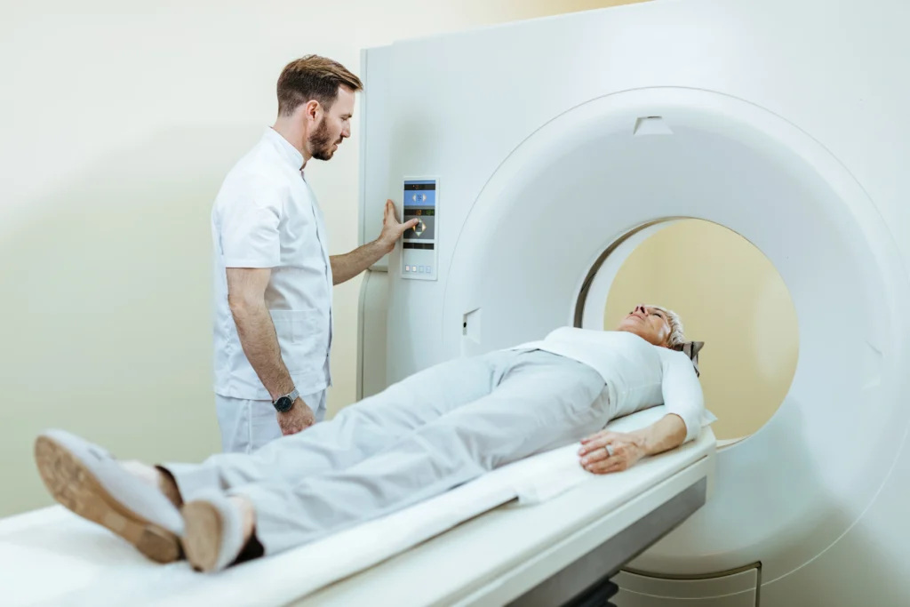
A regular MRI is a basic imaging test. It’s used by doctors to examine various areas of the body. For instance, an MRI can be used to explore the brain, spine, joints, muscles, and numerous organs. The principle is very straightforward. You are on a moving table and it slowly goes into the scanner. Magnetic and radio waves are then used to form detailed images of the machine. You need to stay still during the scan. Otherwise, pictures may be blurred when moving the body. The machine is capable of producing loud knocking noises. So, patients are generally provided with ear plugs or headphones. The follow-up recommended for a moving organ may not be a regular MRI. The exact technique used will depend on the area of the body that is being examined.
How Is Cardiac Magnetic Imaging Different From a Traditional MRI?
The biggest difference is simple: cardiac MRI is designed around a moving heart.
Focus on the heart and its movement.
A regular MRI can take pictures of many body parts. Cardiac MRI focuses on the heart and nearby blood vessels. It can show the heart’s shape and size. More importantly, it can show how the heart moves and pumps blood.
Special images and timing
The heart beats many times during a single scan. That movement can make ordinary pictures unclear. Therefore, cardiac MRI uses heartbeat information to time the images. ECG patches help the scanner know when to capture them. Breath-holding can also reduce movement from the chest. Cardiac MRI can also provide information about the heart muscle itself. For example, doctors may look for scar tissue, inflammation, or other changes. So, the key difference is not the basic MRI machine. The difference lies mainly in how the scan is planned and what doctors want to learn.
What Can Cardiac MRI Detect?
Cardiac MRI can help doctors understand several heart problems. It can show changes in the heart muscle, including damage or scarring. It can also provide information about inflammation and how well the heart pumps. Doctors may use it when they need a closer look at heart muscle disease. It can also help assess some valve problems, blood-flow concerns, and conditions present from birth. For example, another test might show that part of the heart is not working normally. A cardiac MRI can then provide more detail about that area. The benefits of cardiac MRI include detailed pictures of both heart structure and function. However, the test is not necessary for every patient. Your doctor decides based on your symptoms and other test results.
What Happens During a Cardiac MRI?
Before the scan, tell the medical team about any implants or metal inside your body. Also mention previous surgeries, pregnancy, allergies, or kidney problems. These details help the team plan a safe scan. During the Cardiac MRI scan procedure, you usually lie on a comfortable table. Staff place small ECG patches on your chest. The table then moves into the scanner. The machine makes loud sounds while taking pictures. You will receive instructions through headphones. At certain points, you may need to hold your breath briefly. Some scans also need contrast medicine through a small tube in your arm. The scan commonly takes around 30 to 90 minutes, depending on the type of examination.
When Might a Doctor Recommend a Cardiac MRI?
A doctor may recommend cardiac MRI when other tests do not provide enough information. For example, an ECG or echocardiogram may show something that needs a closer look. The scan can help investigate heart muscle problems, unusual heart structure, scarring, inflammation, or certain blood-flow concerns. However, doctors do not order it automatically. They consider your symptoms, medical history, examination, and earlier test results before choosing the right scan.
Conclusion
Cardiac MRI is not simply a regular MRI aimed at the chest. It uses special timing to capture the moving heart more clearly. Therefore, it can provide useful information about heart structure, movement, and muscle health. Uses of heart MRI imaging depend on each patient’s condition and medical needs. If your doctor recommends this test, ask what they want to check. ACE Heart can help you understand your heart care options.
Frequently Asked Questions
Is cardiac MRI painful?
No. The scan itself should not hurt. However, the machine can feel noisy or enclosed.
How long does a cardiac MRI take?
The time varies. Many cardiac MRI examinations take between 30 and 90 minutes.
Can people with pacemakers have an MRI?
Some people can. The medical team must check the device before the scan.
Do all cardiac MRI scans require contrast?
No. Doctors use contrast when they need additional information from the images.
Can cardiac MRI replace an echocardiogram?
Not always. Both tests provide different types of information. Your doctor may recommend one or both.